Spinning Disk W1 Confocal System
Field scanners are renowned for their low dosage, specimen-friendly characteristics, making them ideal for live cell or organism applications.
In vivo microscopy of dynamic processes in cells and organisms requires very fast and sensitive acquisition methods. Confocal laser scanning microscopy (CLSM) is inherently speed-limited by the beam scanning movements. In contrast to single-beam scanning systems, the parallelized approach of multi-beam scanning is much faster. Spinning disk confocal microscopes are therefore very well suited for fast in vivo imaging.
Principle
Spinning disk confocal microscopy
The spinning disk confocal microscopy utilizes multiple pinholes or slits to project a series of 1000 or more parallel excitation light beams onto the specimen in a multiplexed pattern that is subsequently detected after fluorescence emission passes through the same pinholes or slits. The technique is highly useful for high speed imaging of living cells expressing fluorescent proteins or stained with membrane-permeant synthetic dyes. Photobleaching and phototoxicity are reduced with spinning disk microscopy, but a tighter light budget requires very sensitive camera systems for optimum signal detection.
Configuration
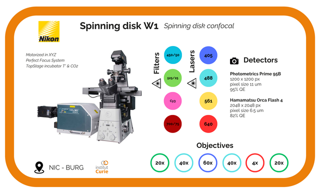
The actual system offers a versatile CSU-W1 with his two disks of 50um and 25um two pinhole-sizes to cover a wider range of objective magnifications. The 25um pinhole size allows for confocal imaging of larger specimens due to its unprecedented lower magnifications and larger fields of view.
The new pinhole design significantly reduces pinhole crosstalk, which is particularly important with thicker specimens. The flexible configuration, with a motorized relay lens for magnification switching, allowing to reply to a large field of applications.
The field of view of the CSU-W1 is nearly 4 times that of the previous CSU-X1, making it ideal for scanning applications of large areas and low magnification imaging of large specimens. This bigger of view is a perfect match for large format sensors. In practice, a wider field of view and higher image quality than previous models, with an enhanced Signal‐to‐Noise ratio, can be acquired
Objectives
The specificity of our set-up is the water immersion objective providing a bigger working distance for an optimal life imaging. It is making possible with a new and essential accessory the water dispenser which replenish automatically the water without any effort during the long time lapse.
Magnification | Type | Immersion | N.A | WD | |
4x | Plan Fluor | Dry | 0.2 | 20 | |
20x | Plan Fluor | Water | 0.95 | 1 | DIC |
40x | Apo Lambda | Water | 1.15 | 0.61-0.59 | DIC |
40x | Apo Lambda LWD | Water | 1.25 | 0.2-0.16 | DIC |
60x | Plan Apo VC | Water | 1.27 | 0.18 – 0.16 | DIC |
Filters in the CSU-W1 Yokogawa
DM | Emission filter | |
Filter Wheel 1 | 404-06/487-89/561-68/633-47 | |
Filter Wheel 2 QuadB | 420-60/510-31/590-624/677-722 | 420-60/510-31/590-624/677-722 |
DAPI | 417-77 | 435-85 |
FITC | 500-50 | 515-55 |
mCherry | 604-79 | 577-630 |
Cy5 | 672-712 | 663-738 |
Filters in widefield illumination
Filters : | Excitation filter | DM | Emission filter |
DAPI | 340/80 | 400 | 435/85 |
FITC | 465/95 | 505 | 515/55 |
mCherry | 542/82 | 601/905 | 604/79 |
Cy5 | 559/950 |
Associated Devices
The current W1 spinning disk is setted two different types of cameras, an EMCCD and a sCMOS camera for different imaging application requiring the sensitivity and/or the resolution.
To response to a fine and precise in depth acquisition, the motorised and triggered piezo stage allows a fast and fine acquisition in a bigger range of depth (500µm).
To maintain the best condition in life imaging, the system is equipped by the last new generation portable stage top incubator offering a large optional accessory ( a special holder design, a microfluidic system, a thermobox…).
Illumination |
Detector |
Software |
Z motor |
T° & CO2 |
EPI illumination : Spectra X, Lumencor |
Flash 4.0 V2 sCMOS camera, Hamamatsu |
NIS-Elements |
Fast-Z motor: Piezo stage Nano z500, Mad City Lab (MCL) |
Stage top incubator STXF-TIZWX-SET Tokai Hit Co. |
Confocal : Oxxius laser (405,488, 561, 640) |
Prim95B EMCCD camera, Photometrics |
|
Techniques
- Brightfield
- Epi-fluorescence
- Time lapse acquisition in 2D, 3D, multiposition and multicolors
- Fast Scan Microscopy (LSM)
Applications
- Tissue, Large mosaic imaging
- Fixed and Lived Cell Imaging