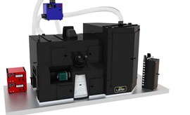
LIPSI Microscope
Nikon Europe has developed, in collaboration with key players in the field Prior scientific and Life Imaging Services, a fully incubated, high content screening platform that enables high speed acquisition for up to 20 well plates. Together with a analysis tools, Nikon enables you to get results from live cells in real time.

Principle
Hight Content Screening HCS
Recent advances in hardware and software innovation for automated image analysis now permits researchers to rapidly screen and analyse hundreds of thousands of images.
High-content screening (HCS), which combines automated fluorescence microscopy with quantitative image analysis, allows the acquisition of unbiased multiparametric data at the single-cell level. This approach has been used to address diverse biological questions, and identify quantitative phenotypes of varying complexity, in numerous different model systems.
In practice, one may acquire, for example, a 96 well plate by scanning the wells faster with a large field of view (up to 25mm). In live imaging, the well plates are hosted in the dedicated opaque chamber to preserve the cells. Without any temperature shock during transfer under a temperature-controlled environment, CO2 and humidity/hygrometry, we can follow the growing cells.
In addition, due to the combination of fluorescence imaging and bright field illumination, the application in immuno fluorescence and histology find a real interest for a large number slice screening.
Configuration
LIPSI utilises all the functionality of the Eclipse Ti2 inverted microscope, including its 25mm field of view and capacity of high speed imaging, while offering modularity and expandability. LIPSI can easily be upgraded with different confocal modalities and accessories to ensure that your platform can evolve with your research needs.
The current configuration of LIPSI is equipped with an LSM microscopy for the high-speed and resolutive confocal A1R HD25 coupled with wide field color camera imaging Nikon DS-Fi3 colour for the histiological acquisitions. Fully automated and motorised from the plate handelling to the stage, we can acquire, visualize and analyse in the real time the data.
Objectives
| Magnification | Type | Immersion | N.A | WD | |
| 4x | Plan Fluor | Dry | 0.2 | 20 | |
| 10x | CFI Plan Fluor | dry | 0.45 | 4 | |
| 20x | CFI Apo LWD Lambda S | dry | 0.95 | 0.99 – 0.9 | DIC |
| 40x | CFI Plan Fluor | dry | 0.95 | 0.21 | - |
| 40x | CFI S Fluor | Water | 1.15 | 0.61 – 0.59 | DIC |
| 60x | CFI Plan Apo VC 60XC | Water | 1.2 | 0.31 – 0.28 | DIC |
Filters
| In widefield illumination | Excitation filter | DM | Emission filter |
| LED-DAPI/FITC/TRICT/Cy5-B | 352-404/461-88/543-66/626-44 | 414-50/500-30/580-611/660-800 | 414-50/500-30/580-611/660-800 |
| LED-CFP/YFP/mCh-A | 426-50/498-520/567-589 | 464-89/532-54/603-800 | 464-86/532-54/603-800 |
| In Confocal illumination | DM1 | DM2 | Emission filter |
| 405/488/561/640 | 495LP | 450/50 | |
| BS 20/80 | 560LP | 525/50 | |
| 640LP | 595-50/700-75 |
Associated Devices
| Illumination | Detector | Software | Z motor | T° & CO2 | Motorization |
| EPI Illumination : Spectra X | Color camera DS-Fi3 | NIS-Elements | Ti2 Z Motor | Special design, Life Imaging Service, LIS | Prior Robot |
| Confocal LU-N4: 405/488/561/640 nm lasers built-in AOTF |
2 GaAsP PMTs + 2 Multi-Alkali PMTs Spectral detector unit with 32 channels |
RaspBerry Camera, LIS |
|
Techniques
- Brightfield
- Fluorescence
- Live Imaging 4D5D : Time lapse acquisition in 2D, 3D, multi-position and multicolors
- Fast Scan Microscopy (LSM)
- Screening
Applications
- Cell imaging, drugs testing
- Life Imaging for organoids
- Fish development
- Histology scanning