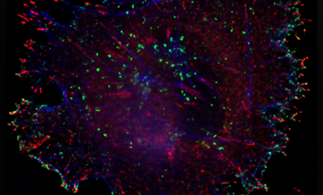
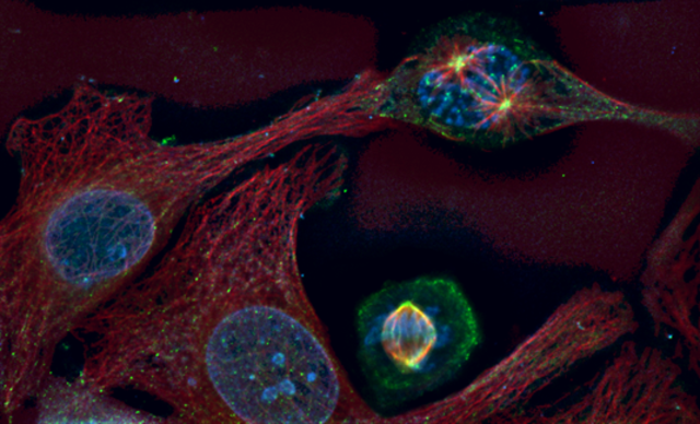
3D Deconvolution Microscope

Fluorescence Microscopy
The absorption and subsequent re-radiation of light by organic and inorganic specimens is typically the result of well-established physical phenomena described as being either fluorescence or phosphorescence. The emission of light through the fluorescence process is nearly simultaneous with the absorption of the excitation light due to a relatively short time delay between photon absorption and emission, ranging usually less than a microsecond in duration. When emission persists longer after the excitation light has been extinguished, the phenomenon is referred to as phosphorescence. More..
Deconvolution
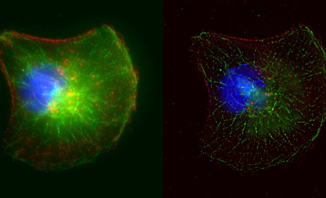
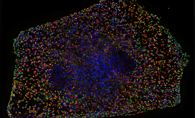
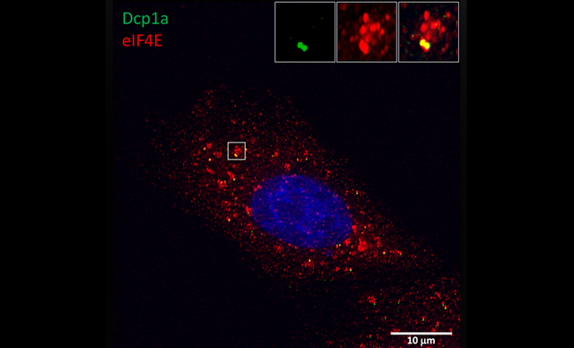
Deconvolution is a computational method that treats the image as an estimate of the true specimen intensity and using an expression for the point spread function performs the mathematical inverse of the imaging process to obtain an improved estimate of the image intensity. MicroscopyU
Configuration
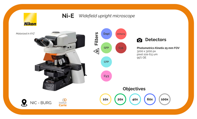
Objectives :
Magnification | Type | Immersion | N.A | WD | DIC |
100x | Plan Apo VC | Oil | 1.4 | 0.13 | Yes |
Filters :
Name | EX | BS | EM |
DAPI | AT350/50x | T400lp | ET460/50m |
CFP | ET436/20x | T455lp | ET480/40m |
GFP | ET470/40x | T495lpxr | ET525/50m |
Cy3 | ET545/25x | T565lpxr | ET605/70m |
mCh | ET560/40x | T585lpxr | ET630/75m |
Cy5 | ET620/60x | T660lpxr | ET700/75m |
Associated Devices :
Illumination | Detector | Software | Z motor | T°C & CO2 |
Intensilight | Photometric CCD Camera CoolSNAP HQ2 | Metamorph | NiE Z motor | NO |
Techniques
-
Brightfield
-
Deconvolution
Applications
- Immunofluorescence fixed samples