Intravital Facility Instrumentation

Instrumentation
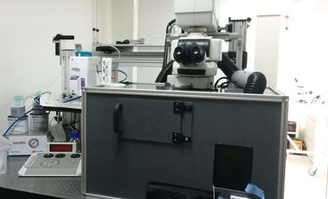
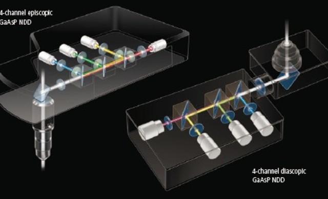
The system is based on an Upright Nikon NiE A1R MP microscope in a special version optimized for wavelengths up to 1300 nm (scanning head and objective). It is equipped with 4 highly sensitive detectors (GaAsP PMT). A specialized motorized stage for animal handling and a piezo z-motor allow fine movements of the areas to be imaged and to gain accuracy when acquiring 3D images. This system can provide structural and functional information with subcellular resolution, sufficient for identifying intracellular organelles and monitoring their trafficking.
Detailled equipment
Upright NiE microscope with A1RMP scanning head
- Conventional and resonant scanner
- Gas detector : Super high-sensitive GaAsP NDD

Spectra-Physics Insight Deepsee laser
- 680-1300 nm, 120 fs pulses
- Automatically maximizes IR laser alignments with a single click, rapid and ideal when working on living animals.
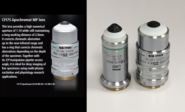
High-NA objectives ideal for multiphoton imaging
Specifically designed for the use of high IR wavelength
- 4x NA 0.22
- 10x NA 0.45
- 16x NA 0.8
- 25x NA 1.1 PlanApo LambdaS

- Luigs&Neumann XY motorized stage
- Microscope incubator
- Mouse monitoring (MouseOx), anesthesia and hydratation systems
Image processing and sharing
Management of microscopy image data is a bottleneck in intravital experiments, specially when considering the masses of data generated from one single in vivo experiment (up to 0.5 Tb per in vivo experiment -one mouse, over 2 to 3 months). We thus provide access to the Image Database “CID-iManage” which provides sharing and browsing of the generated large datasets between distant sites. Moreover, CID can be connected to integrated software packages such 2D and 3D image registration or motion compensation with mobyle-serpico.rennes.inria.fr which are essential issues to be addressed in intravital imaging.

